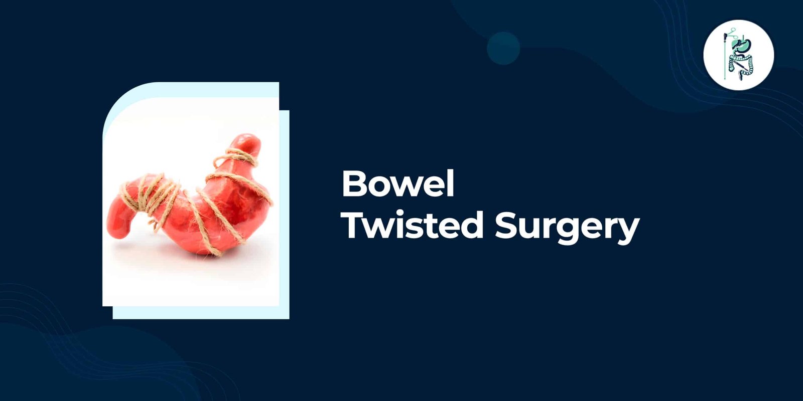
Bowel twisting, also known as volvulus, occurs when the intestine rotates around itself and blocks the flow of digestive contents while cutting off blood supply. This emergency condition often requires prompt surgical intervention to prevent serious complications.
Causes range from congenital malrotation to acquired issues such as adhesions or intussusception. Symptoms may present suddenly with severe pain and vomiting or develop gradually in cases of partial obstruction.
Diagnosis relies on physical examination, X-rays, and CT scans to locate the twist and assess bowel viability. Surgery may involve untwisting the bowel, removing damaged segments, or creating a stoma. Recovery focuses on restoring digestive function and monitoring for complications, with follow-up care essential to prevent recurrence.
Understanding Bowel Twisting (Volvulus)
Volvulus occurs when a segment of intestine twists upon itself, creating a closed loop that traps contents and cuts off blood supply. This condition represents a type of mechanical obstruction where the physical twisting blocks the passage of material through the digestive tract.
Malrotation during fetal development can leave the bowel suspended by a narrow pedicle that permits twisting. The intestine fails to attach properly to the abdominal wall, creating excess mobility that allows the bowel to rotate around its base and form a volvulus.
Functional obstruction differs from volvulus because movement simply stops without any physical twisting present. In functional cases, the bowel loses its ability to contract and propel contents forward, yet no mechanical barrier blocks the passage.
Both types create intestinal obstruction and require prompt medical evaluation. Volvulus demands surgical intervention to untwist the bowel and restore blood flow, while functional obstruction may respond to supportive measures that address underlying causes.
Causes and Risk Factors
Several anatomical and acquired conditions increase the likelihood that the bowel will twist or become blocked. Intestinal obstruction can develop from multiple sources that affect normal movement through the digestive tract. Understanding these factors helps identify patients who may need timely surgical intervention.
Congenital conditions such as malrotation place the intestines in abnormal positions from birth. This arrangement allows the bowel to rotate around its blood supply and form a volvulus. Adults rarely experience this issue unless they had prior symptoms that went untreated.
Acquired causes appear more often in clinical practice. Adhesions remain the most frequent reason for adhesive SBO in adults after previous abdominal procedures. Bands of scar tissue can pull segments of small bowel into fixed positions that promote twisting or blockage.
Additional mechanical issues include intussusception where one segment telescopes into another and gallstone ileus in which a gallstone lodges in the small bowel. Both conditions create closed loop obstructions that often require urgent repair through emergency surgery.
Recognizing Symptoms
Bowel obstruction often announces itself first through pain and visible changes in the abdomen. Early recognition helps patients understand when symptoms require prompt medical attention. Distinguishing between sudden and gradual patterns guides decisions about care.
Acute presentations develop rapidly and demand immediate evaluation. These signs include severe discomfort combined with vomiting and inability to pass stool or gas. Patients may notice dramatic swelling around the umbilicus area.
Chronic presentations appear more gradually and may come and go. Partial blockages often produce recurring discomfort that temporarily improves before returning. Recognition of these patterns helps determine whether evaluation can wait or needs urgency.
Both types relate to conditions like volvulus or malrotation that may require surgical intervention. Understanding symptom progression supports faster decisions about imaging tests or physical examination. Timely identification improves outcomes when intestinal obstruction develops.
Acute Warning Signs
Severe, unrelenting abdominal pain combined with vomiting that turns green signals a possible strangulated loop. These symptoms often indicate complete blockage requiring emergency surgery. Rapid changes demand immediate hospital evaluation rather than scheduled visits.
Cramping discomfort may start intermittently before becoming constant and intense. Bilious vomiting or green vomit frequently accompanies this progression. The abdomen shows rapidly increasing distention as gas and fluid accumulate.
Complete inability to pass stool or gas defines obstipation. This sign distinguishes mechanical obstruction from milder digestive issues. Strangulation threatens bowel tissue when blood supply becomes compromised.
Patients experiencing these combined symptoms should seek emergency care immediately. Conditions like closed loop obstruction or intussusception can progress within hours. Quick action prevents complications such as perforation.
Chronic Indicators
Some patients experience repeated, milder episodes that resolve temporarily before returning. These patterns suggest partial obstruction that has not yet progressed to complete blockage. Monitoring helps track whether symptoms worsen over time.
Intermittent bloating after meals often signals ongoing issues with small bowel passage. Alternating constipation and diarrhea may point to incomplete mechanical obstruction. Recurrent low-grade distention frequently accompanies these digestive changes.
Conditions like adhesive SBO or narrowing from prior adhesions commonly produce these fluctuating symptoms. Functional obstruction may also cause similar intermittent patterns. Patients should note frequency and triggers during episodes.
Persistent or worsening chronic signs warrant scheduling medical evaluation. Imaging tests such as CT scan help identify the underlying cause. Early assessment may prevent progression to acute bowel obstruction requiring urgent repair.
Diagnostic Procedures
Diagnosis begins with a hands-on examination and proceeds to imaging that can locate the site and cause of the blockage. Doctors start by inspecting the abdomen and listening with a stethoscope. They check for high-pitched bowel sounds or complete absence of sounds that signal intestinal obstruction.
Next, patients undergo upright and supine X-rays to reveal air-fluid levels or dilated loops of small bowel. When plain films remain inconclusive, a CT scan with intravenous contrast becomes the preferred tool. This study identifies transition points where the bowel twists or blocks, and it helps distinguish between mechanical obstruction and functional obstruction.
Contrast studies such as small bowel follow-through or enteroclysis provide additional detail when needed. These tests highlight areas of narrowing or twisting that standard imaging might miss. In cases of suspected colonic involvement, lower endoscopy with a sigmoidoscope or proctoscope allows direct visualization of the lower bowel.
Physical examination findings guide the choice of each test. Abdominal pain, vomiting, green vomit, bloating, and abdominal distention prompt urgent evaluation. Early detection of volvulus, malrotation, or closed loop obstruction improves the chance of successful surgical intervention without complications such as strangulation or perforation.
Pre-Surgical Preparation
Before any operative intervention, the care team stabilizes the patient and reduces the risk of anesthesia complications. Medical staff address immediate concerns related to bowel obstruction and intestinal obstruction first. This step prepares the body for general anesthesia during twisted bowel surgery.
Placement of a nasogastric tube helps relieve pressure from the stomach and upper digestive tract. The tube allows continuous suction to remove trapped air and fluid. This action reduces vomiting and eases abdominal distention before surgery begins.
Intravenous fluid resuscitation follows with careful monitoring of electrolytes. Doctors correct imbalances that develop from vomiting and reduced intake. Blood typing and cross-matching occur at the same time to prepare for possible transfusion during the procedure.
A urinary catheter is inserted to track urine output throughout the operation. This measurement helps the surgical team monitor kidney function and fluid balance. Accurate tracking supports safe management under general anesthesia for bowel resection or other repairs.
Surgical Treatment Options
The choice of operation depends on whether the twisted bowel remains viable and how much length is affected. Emergency surgery becomes necessary when patients show signs of perforation or ischemia that threaten life.
Intestinal obstruction from volvulus requires prompt surgical intervention once imaging tests confirm the diagnosis. Surgeons evaluate bowel color, movement, and blood supply before deciding between simple untwisting or removal of damaged segments.
Viable tissue allows for detorsion alone or with fixation procedures. Irreversible damage from strangulation forces resection of the affected area followed by reconnection or diversion.
General anesthesia supports these operations while teams monitor for complications such as anastomotic leak or paralytic ileus during recovery. Early action reduces risks tied to complete obstruction and closed loop patterns.
Detorsion Techniques
When the bowel is still healthy, surgeons untwist the loop and may anchor it to prevent recurrence. Access occurs through midline laparotomy or laparoscopy depending on patient stability and anatomy.
After manual detorsion, cecopexy secures the cecum to the abdominal wall using sutures. Children with malrotation often receive a Ladd procedure that divides peritoneal bands and widens the mesenteric base.
Persistent distention may prompt addition of cecostomy for ongoing decompression. These fixation steps address mechanical obstruction and lower chances of future twisting episodes.
Surgeons also perform concurrent lysis of adhesions when present. This combination reduces adhesive SBO risk and supports normal bowel function after the initial repair.
Resection and Anastomosis
Dead or severely damaged bowel must be removed and the healthy ends rejoined. Assessment relies on color, peristalsis, and Doppler signals to confirm viable tissue before any cutting begins.
Surgeons resect the non-viable segment and perform primary anastomosis for small bowel cases. When reconnection poses too much risk, creation of an ileostomy or colostomy protects the patient during healing.
Lysis of adhesions occurs at the same time to prevent future episodes of adhesive SBO. This step addresses scar tissue that commonly contributes to recurrent bowel obstruction after prior abdominal operations.
Postoperative monitoring focuses on signs of anastomotic leak and electrolyte imbalance. Most patients regain bowel sounds within days when fluid resuscitation was adequate before and during the procedure.
Post-Operative Care
Immediate recovery focuses on restoring gut function while watching for early surgical complications. Patients remain under close observation after bowel twisted surgery to ensure the digestive tract begins working again safely. Monitoring helps detect issues before they worsen.
The nasogastric tube stays in place until bowel sounds return. This device prevents stomach contents from building up during the initial healing phase. Removal happens only when the medical team confirms normal gut activity through physical examination.
Diet advances slowly from ice chips to clear liquids once the patient tolerates fluids without nausea. Intravenous fluids continue throughout this period to maintain hydration and correct any electrolyte imbalance. Gradual progression reduces strain on the surgical site.
Daily wound inspection checks for signs of anastomotic leak or infection at the repair location. Early ambulation begins as soon as possible to prevent paralytic ileus and reduce thromboembolism risk. Staff assist patients with short walks several times each day.
Recovery Timeline and Expectations
Return of bowel activity and full strength varies with the extent of surgery and any coexisting illness. Patients who undergo repair for twisted bowel or volvulus often experience different healing speeds depending on whether a bowel resection or stoma creation occurred during the procedure.
Days 1 through 3 after emergency surgery typically involve an nasogastric tube and intravenous fluids. These measures help manage paralytic ileus and support recovery from general anesthesia while the digestive tract remains inactive.
Between days 4 and 7 most patients transition to oral intake once bowel sounds return. Discharge planning begins during this window, and care teams assess readiness for home based on tolerance of liquids and pain control.
At home during weeks 2 through 4, lifting remains restricted to protect healing tissues. Follow up imaging may occur if a colostomy or ileostomy was created to monitor for complications such as anastomotic leak or early adhesions.
Months 2 and 3 focus on gradual return to normal diet and daily movement. Patients watch for signs of recurrent obstruction including abdominal pain, vomiting, or distention and report these changes promptly to their surgical team.
Potential Complications
Even successful operations carry risks that patients should recognize early. Intestinal obstruction surgery for conditions such as volvulus or malrotation requires careful monitoring afterward. Patients need awareness of warning signs that may develop during recovery.
Anastomotic leak represents one serious concern after bowel resection. This issue often presents as fever combined with increasing abdominal pain. Prompt reporting of these symptoms allows medical teams to intervene before further damage occurs.
Prolonged paralytic ileus can delay return to normal eating. This condition slows digestive function and may extend hospital stays. Patients experiencing persistent nausea or inability to tolerate oral intake should alert their care team immediately.
Additional issues include recurrent obstruction from new adhesions and problems related to ostomy creation. Ostomy-related issues such as skin irritation or high-output losses often require specific dietary adjustments. Early recognition of worsening symptoms improves overall outcomes after surgical intervention for bowel obstruction.

Dr Vishant Deo Editorial Team
Led by Dr. Vishant Deo (MBBS, MS General Surgery), one of the best Laparoscopic & Cancer Surgeons in Siliguri.
A gold medalist in MBBS, Dr. Deo has trained at top institutions including Tata Memorial Hospital, AIIMS, and Fortis Hospital. He is an active member of the Association of Surgeons of India (ASI) and practices at Star Hospital, Siliguri.
All content is reviewed for medical accuracy and patient education.